A 'floating' knee meniscus is really a radiological term used when the doctor reading an MRI scan notices in certain views that there appears to be fluid above and below the meniscus, so that it is no longer in close communication to the flat top of the tibia, upon which it usually sits.
To understand this, one needs to know that the middle part (body) of the meniscus has ligament attachments both to the femur above and to the tibia below.
 |
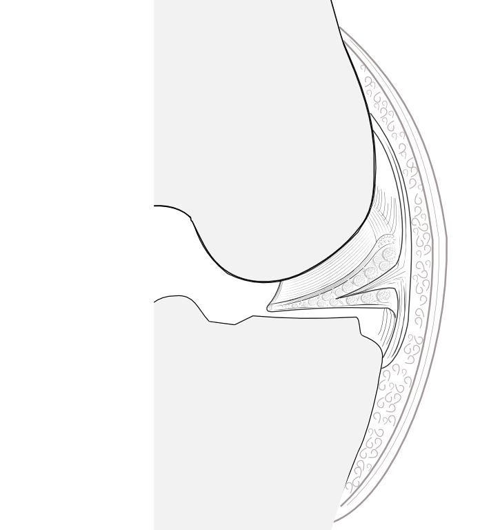
On the left of my illustration is the way most illustrators depict the meniscus - just sitting on the top (plateau) of the tibia bone. I generally draw it this way as well, for simplicity. But the anatomy is rather more like you can see on the right - where the blue arrow is - the meniscus connects to the waterproof capsule that encloses the knee joint, and fibrous strands go from the meniscus to the bones above and below the joint, tethering it down and limiting its movement. My illustration still needs some amending, because actually these 'menisco-tibial' and 'menisco-femoral' ligaments are rather shorter, and attach closer to the joint line than I have shown here. |
Although this is just a rough sketch, this next picture on the left perhaps shows the normal situation a bit better better.
A violent injury, perhaps involving twisting, may tear that ligament attachment off the tibia bone.
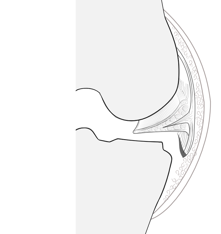
See the sketch on the right. The meniscus itself may be undamaged, but now this ligament 'anchor' has been pulled right off. This is called an 'avulsion'. Because of the other injuries that may occur at the same time, the avulsion may be missed, but MRI scan may hint at it because of the appearance of an unusual amount of joint fluid under the meniscus, making it appear as if it is floating.
As the patient takes weight on the leg, the meniscus may 'extrude' - push outwards - and fails in its normal duty to properly distribute the forces going through the joint.
Further reading/watching:
1. Dr Paletta's presentation, courtesy of Arthrex
2. The “Floating” Meniscus: MRI in Knee Trauma and Implications for Surgery
