It is not possible to understand about knee flexibility without a good working understanding of the anatomy of the knee joint, the knee cavity and the tissue folds lining the joint.
 First published in 2017, and reviewed August 2023 by Dr Sheila Strover (Clinical Editor)
The Anatomy of Knee Flexibility - course
- Introduction to the course on the Anatomy of Knee Flexibility
- Normal knee range-of-motion in men and women
- The joint capsule – why it is key to understanding so much about the knee
- Important structures inside the capsule – & their contribution to knee movement
By the end of this Section, you will understand what has motivated your instructor to build a course like this.
You will take your own first steps to accepting that knee anatomy is central to taking informed responsibility for what happens to your own knee during the rehabilitation process.
This course will describe to you those structures that allow a smooth and full motion of the knee, and particularly the soft tissue structures such as the joint capsule surrounding the cavity of the knee joint and the structures intimately related to the kneecap.
At the end of this course, you should be able to:
- understand the key role of the joint capsule, and how it both contains the internal structures and lubricates the process of joint movement
- see how important the patella is in relation to knee movement, and how intimately it is associated with the quadriceps muscle, and the tendons above and below it
- have an idea about the anatomy of the fat pad and the anterior interval
- realise that the cruciate ligaments both allow movement and contain movement
- know where the menisci are and what their role is with regard to knee movement and knee stability
Foreword
It's a curious truth that I did not enjoy anatomy when I was a student, despite majoring in the subject. And now I am fascinated by the anatomy of the knee. But then again I loathed history at school and now I avidly follow history and archaeology programmes on my computer. Subjects have this way of getting interesting when you want to understand things better....
Twenty years ago I was working with my then husband on a project to help train young surgeons in keyhole surgery (arthroscopy) of the knee and shoulder. We were trying to develop a three dimensional model that could be filled with water to emulate the conditions during arthroscopy, so that they could learn not only the anatomy but also the tricky business of controlling the fluid input and output inside the pressurised joint cavity while trying to perform challenging surgical procedures. Angus did something rather interesting to try and help me - he went into the anatomy department, and injected under pressure the knee joint of a cadaver with some liquid resin that had just been mixed with an accelerator, and waited for the resin to set.
Then when it was solid, he dissected out and handed to me what was an accurate representation of the space within the knee joint! I was amazed - despite being a qualified doctor with an anatomy BSc I had never imagined that the joint cavity was so large and so complex. Suddenly my project took on new life as I understood that the model I was trying to make by fitting two bones together, actually had little to do with the bones and a lot to do with the space between and around them, contained by the soft tissues.
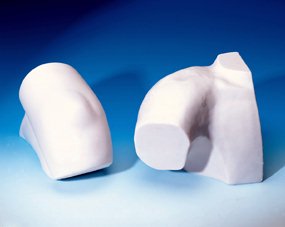 Just for interest, this is a photograph of the final production models (no longer being manufactured - but used on courses for more than a decade).
This concept of a water-tight cavity is key to understanding the interior of the joint, and of course surgeons utilise that when they blow the knee up with fluid during arthroscopy. But it also means that it is a big cavity that can fill up with blood, too, and get inflamed, and infected and scarred up!
INTRODUCTION TO COURSE: The Anatomy of Knee Flexibility - course
NEXT PART: Normal knee range-of-motion in men and women
