Focus on post-operative rehabilitation is critical in managing the patient after surgery for knee arthrofibrosis.
 First published by Dr Noyes in 2008, and reviewed August 2023 by Dr Sheila Strover (Clinical Editor)
- Established arthrofibrosis - options for management
- Arthroscopic surgery (for arthrofibrosis)
- Open debridement and soft tissue release
- Rehabilitation After Operative Procedures for Arthrofibrosis
- Surgery for chronic patella infera
- The tragedy of arthrofibrosis
- Preventing arthrofibrosis
The program of physical therapy following any of the surgical procedures discussed in Parts 7 or 8 of this course is critical in maintaining the knee motion gained from the operation.
We have seen unfortunately many patients who have undergone an operation elsewhere which may have initially been successful in regaining more normal knee motion, but because the rehabilitation was ineffective, the patient soon lost flexion and extension and was right back where they started from before the operation. This frustrating cycle of ineffective treatment is due to either a complete lack of physical therapy after the operation, or to what I believe are insufficient attempts in maintaining motion, such as only using a continuous passive motion machine for many hours each day.
In-Patient Physical Therapy Program
A continuous epidural anesthetic or femoral nerve block, along with appropriate analgesics, is used for 3 to 4 days following surgery. The epidural is regulated to block pain and sensory input, but retains muscle motor control to allow active knee motion and the overpressure program to be described. The block is closely supervised by an anesthesiologist for levels and medication dosage. Nursing personnel performs a skin care and protection program. The block is maintained for a minimum of 72 hours in order to allow a gradual overpressure program to stretch contracted soft tissues.
We follow several measures in order to avoid deep venous thrombosis and pulmonary embolism. First, a careful analysis is done to detect patients at increased risk, including diabetics, obese individuals, or those with venous insufficiency, prior history of phlebitis, or venous clots or embolism. These patients are not appropriate candidates for the epidural program due to the associated inactivity and bed rest. When the epidural is in place, the patient remains mobile and is encouraged to be out of the bed intermittently throughout the day. Anti-embolism stockings are worn and ankle pumps are done for 5 to 10 minutes hourly. The extremity is monitored daily and a calf venous compression device is used. Aspirin (600 mg daily) is prescribed. The patient is monitored for calf tenderness or leg edema and an ultrasound examination of both lower extremities is obtained if any suggestion of a DVP exists.
The patient is taught before the operation how to perform the overpressure exercises. I recommend this type of active range of motion program, which is performed by the patient, instead of a continuous passive motion machine. The amount of tension applied to the foot-calf stocking for achieving knee flexion is controlled by the patient. It is important that other individuals such as the family not assist in the overpressure flexion, as this would involve a flexion manipulation by untrained personnel.
The overpressure extension and flexion exercises are shown in the Figures below.
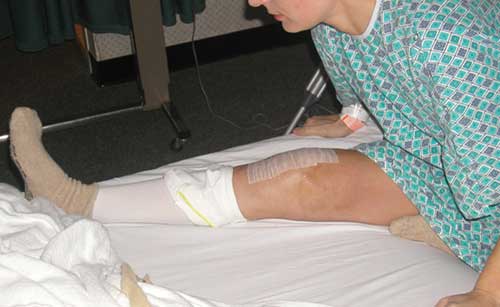
The extension exercise is done first.
The amount of weight to be used is determined before the operation. 15-20-25 pounds of weight is placed over the knee with the foot and ankle supported.
The amount of weight is increased as tolerated. The lowest amount of weight is used to achieve 3° hyperextension at the 3rd day.
[Noyes FR, Barber-Westin SD: Chapter 41: Prevention and treatment of knee arthrofibrosis. This article was published in Noyes’ Knee Disorders: Surgery, Rehabilitation, Clinical Outcomes, Noyes FR, Barber-Westin SD (eds.), pp. 1053-1095, Copyright Saunders, 2009.]
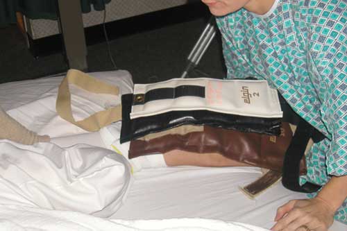
Then, the flexion exercise is done. The affected heel height is marked on opposite thigh before the epidural is begun to determine extent of contracture and starting point. The amount of flexion gained over the first 24 hours is limited to prevent muscle and tissue tearing. Patellar mobilizations are done to avoid patella overpressure with knee flexion.

The patient is placed in a “figure-four” position in bed. A strap or towel is placed, with approximately 15 to 20 pounds of pull allowed on the ankle. The heel-thigh mark is monitored on the opposite thigh to determine gains in flexion.
The flexion overpressure program is done every 30 minutes for 5 minutes while the patient is awake. The goal is to achieve heel-height above patella. I perform a flexion overpressure test to determine whether soft or hard resistance is present and measure the knee flexion angle daily.
The patient performs leg lifts and isometric exercises while in the hospital. It is imperative to control soft tissue inflammation and avoid a hemarthrosis. If soft tissue swelling persists, or thigh muscle pain occurs due to excessive stretching, a nonsteroidal or steroidal dose pack should be considered. After the epidural, I routinely obtain a venous ultrasound to exclude venous clot disease.
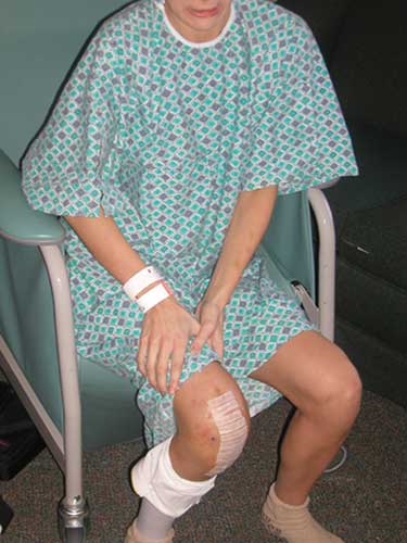
Upon completion of the in-patient program, the exercises must be continued at home 8 times a day. Close supervision by the physical therapist is required, as the gains in knee flexion and extension may be reduced rapidly if the regimen is not maintained, or if pain and swelling prohibit the patient’s ability to perform the exercises. Other exercises to regain normal gait, muscle strength, and flexibility are added under the physical therapist’s guidance. Chronic muscle wasting is a frequent problem in patients with established arthrofibrosis, and it is important to work the quadriceps, hamstrings, and hip muscles as tolerated. Modalities such as electrical muscle stimulation and biofeedback are useful in the early stages of rehabilitation. The muscle stimulator can be used while the patient is performing leg lifts to further enhance the effectiveness of this exercise. Gradually, weight bearing exercises may be incorporated such as wall sits (where the patient sits against the wall with the knees slightly flexed for as long as possible) and balancing on unstable surfaces (such as foam rolls). Swimming and bicycling (with little to no resistance) are excellent, if tolerated by the patient. An experienced physical therapist can progress the program in a safe and effective manner.
PREVIOUS PART: Open debridement and soft tissue release
NEXT PART: Surgery for chronic patella infera
Dr Frank R Noyes

DR NOYES HAS RETIRED.
Based at the Cincinnati Sportsmedicine and Orthopaedic Center in the USA, Dr Frank Noyes is one of the world's most prominent figures when it comes to knee surgery. A prolific researcher and writer, he has published over 200 studies and articles in the world's top...read more