Dr Angus Strover explains the steps a knee surgeon will take in performing an arthroscopy.
 First published by knee surgeon Angus Strover in 2008, and reviewed August 2023 by Dr Sheila Strover (Clinical Editor)
Diagnostic Arthroscopy - The Importance of Method
Hello and welcome to my course on a 'method of routine diagnostic arthroscopy'.
This is an important subject.
For over twenty years now and together with a number of dedicated colleagues, I have been trying to spread the use of a systematic method to the hundreds of young surgeons who attend our skills workshops at The Knee Foundation.
This course is aimed at an 'intermediate' level, and may be of interest to both patients, junior doctors and operating theatre staff.
This is what we are going to cover during the course:
- Firstly, I am going to take you through some steps the surgeon needs to take on the day surgery is decided.
- Then we will skip to the short period before surgery, when the patient still has a chance for last minute interaction with the surgical team.
- There will be a brief discussion of the anaesthetic choices, and the preparation of the patient before the first portal (cut) is made.
- I will run over the anatomy with you.
- I will explain the limitations of some of the instrumentation an arthroscopic surgeon is obliged to use.
- After this I will explain why it is very important that the surgeon should follow a routine in fully examining the knee. I will use pictures and videos to illustrate this section.
- Then a round-up of some of the conditions most likely to be missed by failing to use a systematic approach.
Note that, except for a brief overview in the final lesson, we are not going to talk about the surgical procedures which a surgeon might perform during an arthroscopy - this is the stuff of more complex courses. The purpose of this current course is to show how important method is in arthroscopy - and how one can miss the diagnosis by using faulty method.
Informed Consent
In my practice, I have a really useful three-dimensional model of the knee, and I take every patient through their procedure using this model. It is made by Adam Rouilly and I have found it over the years to be virtually indestructible. This little model is so useful that I give one to each of my departing knee fellows (trainees) when they head off back to their own countries to set up practices there!
The model allows a discussion of various knee cap problems such as tilt and maltracking; it helps to demonstrate the effect of procedures to improve the mechanics of the knee cap; the common sites of arthritis can be identified and relevant procedures for arthritis can be discussed; the cruciate ligaments are easily identified, and the procedures for reconstruction can be explained. And so on.
I think that this model is much more useful than pictures, as the two-dimensional nature of an illustration may cause the patient some confusion - whereas the model allows for a much more sophisticated level of dialogue altogether.
Procedures just before the anaesthesia
The period just before the patient receives the anaesthetic is an important time, and again here a surgeon can miss an important opportunity to -
- reassure the patient that it is he who will be doing the procedure, not another doctor the patient does not know
- check that relevant x-rays have arrived, together with the patient notes
- confirm the procedure which has been signed for, and that the symptoms are still the same
- make sure the SURGEON HIMSELF, under the watchful eye of the awake patient, marks the correct knee with a thick surgical marking pen which won't wash off
Picking up that last point, how easy do you think it is to do an arthroscopy on the wrong knee?
Yes. Terrifyingly easy.
Firstly, because the two knees might both look perfectly normal at the time, as there may be no swelling or external sign of anything wrong with the knee.
The patient is asleep when the surgeon is scrubbing. Also the nurse is draping the knee while the surgeon scrubs, and the surgeon generally accepts that the nurse won't made a mistake!
But a big black arrow on the bad knee, put there by the surgeon himself before the patient is put to sleep, ensures that such a disaster does not happen.
I won't go into detail about the anaesthetic, or discuss whether one should have this or that anaesthetic, but I would just like to tell you about one or two steps which I believe make a great difference to the patient.
I had the good fortune to work closely with a team of anaesthetists (anaesthesiologists) who were expert at doing what is known as 'regional blocks'.
A regional block is a nerve block with local anaesthetic, which blocks the nerve in its lower distribution, in contrast with a local block, which paralyses the nerve structures just around the injection itself. Some anaesthetists (anaesthesiologists) are good at this and are able to produce complete loss of sensation in the knee.
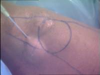
For ordinary arthroscopic procedures we used this for some time, but found that many patients were frustrated that they could not use their quadriceps muscles for sometimes several days afterwards.We now use local anaesthetic in the skin at the sites of the portals before the operation starts and also leave some local anaesthetic in the joint at the end of the procedure.
Here you can see the bulge in the skin where the local anaesthetic is being injected.
Don't worry about those pen marks. You will understand what they are when you get to a later section of the course.
Use of a tourniquet
A tourniquet is an inflateable wrap, which goes around the top of the thigh and gets pumped up to stop the blood flowing to the limb.
Probably the majority of knee surgeons apply and inflate a tourniquet before commencing the arthroscopy, and in addition the scrub nurse usually first exsanguinates the leg (squeezes the blood out using an elastic bandage). This offers the surgeon an advantage in there being virtually no bleeding during the procedure, but I believe this to be a two-edged sword for the following reasons:
- little blood vessels (even small pumping arteries) may continue to bleed after the operation, when any swelling is hidden by the bandages and any pain due to the swelling is attributed to the surgery itself
- there is an increased risk of suffering a clot in the leg after surgery if a tourniquet is used (deep vein thrombosis)
For a routine arthroscopy I do not even apply a tourniquet!
Instead I make a point of controlling bleeding during the arthroscopy by these methods:
- I carefully control the inflow and outflow of fluid into the joint using an irrigation pump, which is ideal. I find this important because any excessive outflow of the irrigation fluid will cause a drop in the fluid pressure inside the joint and will result in immediate bleeding. I usually set the irrigation pump pressure at 50 mm Hg (mercury pressure) and increase this up to 70 if necessary.
- The inflow of fluid into the joint needs to be unimpeded. This means that the diameter of the arthroscope must be large enough to allow a quick flow of fluid if one is going to pass the fluid into the joint via the arthroscope itself. (We'll discuss the arthroscope later.) Most arthroscope suppliers these days provide diagnostic and operative instruments. The operative one allows a sufficiently high flow of fluid into the joint to maintain pressure whilst doing most arthroscopic procedures. The diagnostic one is slimmer, but is not adequate if one is going one to use power instrumentation (like a rotating burr) during any subsequent arthroscopic surgical procedure. The alternative is to use in inflow cannula - a separate wide-bore needle just for fluid inflow.
- I also use adrenaline (epinephrine) in my irrigation fluid in a concentration of 1 part per two million (1:2 000 000). This constricts the smaller blood vessels (capillaries) and is effective in reducing capillary bleeding.
- If there is any arterial bleeding during the procedure, I control this with diathermy (cautery), burning the bleeding vessels to seal them.
For beginners in this technique of arthroscopy it may be practical to apply a tourniquet but not inflate it. It can then be used if necessary. I started this way and graduated to the position of no tourniquet after a period of three or four years.
OK. This is the end of Part 1. In Part 2 I will discuss the basic instrumentation and its limitations.