Plical folds of the lining of the knee joint are often dismissed causes of knee pain.
 First published by knee surgeon Angus Strover in 2008, and reviewed August 2023 by Dr Sheila Strover (Clinical Editor)
As we continue on this arthroscopy voyage together, I just want to touch upon the subject of plicae.
We mentioned them in the anatomy section as folds of joint lining left over from the embryonic stage of knee development and I will go into more detail in the next part of the course. For the moment I want to show you three illustrations to keep the topic started.
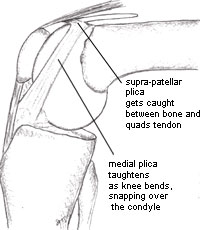
The commonest problematic plica is the medial plica. In the left image it is seen draped like a curtain over the medial condyle, attaching at the bottom to the fat pad, at the top to the wall of the supra-patellar pouch and on the outer aspect to the rest of the joint lining. This last is not appreciated easily from an illustration.
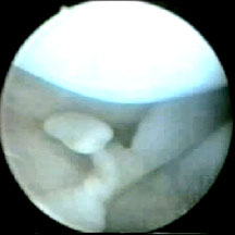
In the next two images, you will see how the plicae can get irritated or damaged when the knee is bent, particularly if they have recently taken a bump and become swollen or thickened.
 |
 |
 |
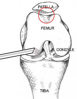
Before swinging the arthroscope up and around the condyles, noting any plicae as we go, the 'gutters' to the side of the joint cavity must be examined. These are often forgotten by the novice, but are notorious for hiding 'loose bodies' - bits of joint surface which have detached and which grow bigger in the joint cavity.
After examining the lateral gutters, the arthroscope is brought up and around the condyle to view the trochlea and the undersurface of the patella - the region known as the patello-femoral joint. Again, this will be dealt with in the next lesson, when we view this region from a new portal.
We are nearly finished this lesson now. I'll just show you a video of this region being examined with the probe. You will notice some abnormal softness of the joint surfaces.
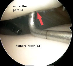
The red circle in the left hand image explains where the probe is probing. Just ignore the arthroscope in the picture - I need to edit the image. In the right image the probe has reached underneath the patella, where the cartilage is not normal. The probe is able to make a dimple (this softening is called 'chondromalacia'). You can see the fluid inlet in the distance.
This is the end of this lesson now. In Part 8 I am going to be telling you some professional secrets!