This case is of a post-traumatic realignment osteotomy.
 First published 2010, and reviewed August 2023 by Dr Sheila Strover (Clinical Editor)
Realignment osteotomy for knee pain - course
Part 1 - Introduction to the subject of knee osteotomy
Part 2 - Osteotomy for varus and valgus deformity
- Indications for varus and valgus osteotomy
- Living with painful varus and valgus deformity
- High tibial osteotomy and distal femoral osteotomy
- Case study of high tibial osteotomy aiding ligament instability
- Benefits of varus/valgus osteotomy
- Potential problems with varus/valgus osteotomy
- Recovery post knee osteotomy
Part 3 - Osteotomy for patellar instability
This was a challenging case of a 17 year boy who came off his pizza delivery bike and fractured his femur.
I am presenting this case as just a very good example of a young patient with major trauma related knee ligament problem, in whom osteotomy surgery has played a key role in correcting the ligament instability.
When he came off his bike, he broke his femur and also injured his knee thereby rupturing the lateral ligaments on the outside and the back of the knee (posterolateral corner - PLC). He also injured his posterior cruciate ligament (PCL) which he avulsed-off which means he pulled the PCL off its attachment at the back of the tibia together with a small piece of bone.
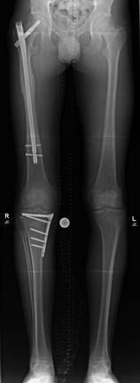
So we carried out quite a complex operation. Firstly, we fixed his femur with a long 'nail' - you can see that on the left side (your left) of this long-leg X-ray, Then we turned him onto his tummy and opened up the back of his knee. Here we located the bits of bone still attached to the PCL and put them back on again (which is called a primary repair of the ligament). Then turned him onto his side and repaired all the ligaments on the outside.
However I wasn’t happy with the PCL repair so once he recovered from his femoral fracture, I took him back to theatre and carried out an osteotomy to valg-ise him. At the same time I increased his tibial slope in order to improve his PCL deficiency and resultant knee instability. I was expecting to tee him up for a revision PCL reconstruction, but as is so often the case, by increasing his slope in a sagittal plane and making it steeper, we corrected the fact that his PCL is not that great and on his follow-up appointment he was very happy with the results of his surgery and was not complaining of any instability.
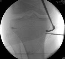
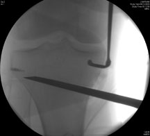
This picture on the left shows the fixation with an anchor on the side to put his ligaments back on again. As part of the osteotomy you can see the drill in position on the inner side of the leg, and that is aiming almost at the fibula. The next image on the right shows the chisel going in under X-ray guidance – the wire is the drill bit and then the slightly flat looking thing is the chisel.
Professor Adrian Wilson

Mr Adrian Wilson is a consultant knee surgeon based in London in the UK. His practice involves all aspects...read more