Recuperation and rehabilitation after knee osteotomy is usually straightforward.
The rehabilitation programme will change as you progress from hospital bed to full recovery.
Hospital phase
The operation takes around 1 hour. Most patients remain in hospital for 3-4 days.
Most patients come in to hospital on the day of surgery. They are sometimes given calming medications prior to surgery. For most patients we use a spinal anaesthetic (as opposed a general anaesthetic), which numbs both legs completely. That lasts for up to 6 hours afterwards and provides pain relief for a number of hours after the operation. We don’t tend to use general anaesthetics (GA) for osteotomy surgery. Some patients are not keen on the idea of this initially, but my anaesthetist is very good at explaining the options to them. He says “Let’s get you down to theatres and get the spinal anaesthetic in. If you then want me to sedate you, you can drift off and if you are OK with it we will keep you awake”. Most patients feel a bit woozy when they go into theatre but then tend to wake to the point where they are a bit more alert, and actually they find that they don’t feel nervous or worried and often end up chatting with the anaesthetist during the procedure.
This is a big procedure and what we don’t want is for our patients to experience unnecessary discomfort or pain. So by having a spinal anaesthetic they wake up completely pain free. To enhance post-operative pain relief, during the operation we inject a lot of dilute local anaesthetic at different stages of the procedure – all around the site of the osteotomy and the surrounding structures. We don’t use a wound drain in this operation, as bleeding is rarely a problem. The local anaesthetic works until the next day – as it wears off, the patient would already have been started on very strong long-acting painkillers. Most patients are comfortable on these tablet painkillers and don’t require any further pain relief, such as a PCA (patient-controlled analgesia). So the combination of spinal anaesthesia, local anaesthetic infiltration (at the time of surgery) and strong pain killers allows the patient to be comfortable even at the early post-operative stage.
If it is very straightforward, surgery from start to finish only takes an hour. If the patient is also having a ligament or an osteotomy is also required on the femur, the total operating time is usually around two hours. It seems like a lot longer to most patients. This is because the the patients spend a bit of time having their anaesthetic before the surgery, and after the surgery they are taken to recovery for an hour or so until they are ready to go back to the ward.
After the operation, the patient has a TED compression stocking applied to the operated leg, to both protect against blood clots (DVTs and PEs) and to reduce swelling after the operation. Obviously the bone has been cut and opened and can potentially bleed, and we want the wound to heal up, so we have found the compression stocking helps a lot with swelling afterwards.
We also use a cryo-cuff which is a special device that is velcroed around the knee and helps to reduce swelling. It is filled with water and ice and cools the knee. It stays on for 20 minutes and then comes off and regular use of the cryocuff for 2, 3, 4, perhaps 5 days that you are in hospital depending on how big the operation was.
In addition, special pump (AV) boots are placed on both legs. They are taken off when the patient gets up and walks around. They markedly reduce the chance of blood clots forming in the legs after surgery. As mentioned before, the patient will also have a TED stocking on the leg they had the operations on (to reduce swelling), and the cryocuff is also on when the patient is in bed. The AV boot is just a little ankle device that velcros around the ankle – the pneumatic bit is in the sole – so every few seconds it compresses the blood in the sole of your foot and sort of tickles your feet.
Patients start mobilising (with their physiotherapists' help) the day after the operation, initially with the aid of 2 crutches. You CAN fully weight bear most of the time because the plate we use is so strong, but by not weight-bearing fully for the first two weeks, the wound is allowed to heal. So prior to discharge, the goal is to be safe mobilising on two crutches, ie. walk and manage a flight of stairs safely, and have good pain control with minimal/limited swelling. Most of the patients who have this operation on a Thursday, are home by Monday and reasonably comfortable.
The reason we don't tend to use an epidural anaesthetic for this type of operation is that anaesthetists have moved away from epidurals in the UK, as the epidural is slightly more difficult to manage because it is a continuous technique, the idea being that you leave in the epidural catheter in and then it gets topped up with anaesthetic solution. Nurses have to be specially trained to manage them and there is an increased risk of infection as the catheter communicates with the spine. One-off spinal injection is the way we are now going in the UK with regional anaesthesia and we don’t tend to use epidurals for anything any more in orthopaedics. What has been revolutionary though is a technique that has come from Australia - which is injecting very large volume of dilute local anaesthetic agent in and around the knee, during the operation. They do it for knee replacements as well and it is amazingly effective. By injecting the local anaesthetic all around the operation site and allowing the tissues to really soak it up, the patient can be virtually pain-free when the operation is completed. We tend to use a local anaesthetic called Marcaine which lasts up to 14 hours. We don’t tend to do regional nerve blocks alone for osteotomy surgery. It would either be a spinal OR femoral nerve block PLUS this local anaesthetic infiltration, and most patients get the spinal.
One of the risks associated with having a spinal is that you may not be able to pass urine in the evening on the day of the surgery so you may need to have a temporary catheter. Because the anaesthetists have refined their technique over the last 5-10 years, they have become very good at ‘taking out’ the sensory nerves – pain nerves – and preserving motor nerves, which makes urinary retention, ie, inability to pass urine, less likely. You can quite frequently actually move your leg after having had the spinal injection, with good motor control so that patients frequently don’t wake up with a leg that they can’t move. Not blocking the motor nerves also means that the ones supplying the bladder are spared making it easier to pass urine. So that is something that has moved on a lot in recent years. And although having a catheter put in is an inconvenience, it is rare disadvantage of this brilliant pain-relieving technique.
Recuperation during first few weeks
Patients go home on crutches, with no need for a splint or knee brace. They need to picked up by a relative as they wont be able to drive initially. Patients are given strict instructions to do very little for the first six weeks, just some simple exercises to stimulate and build the quadriceps muscles, and do some straight leg raising (lifting it straight off the bed). That is about it in terms of the first few weeks. Then they build up to bending the knee, and aim to progress their bend to 90 degrees.
Out-patient physiotherapy commences a week following surgery. They get quite intensive physiotherapy as an inpatient, and then an appointment is made so that within that first week following discharge, the patient is seen by their physiotherapy. They will then ideally go on to have twice weekly physiotherapy for the first six weeks.
During those six weeks you progress from two crutches down to one crutch down to a stick down to nothing but most people by six weeks are able to walk without any walking aid or at most just a stick. For someone who has got a sedentary office type job and is motivated, they can get back to that type of work at the six week stage. This is much, much sooner than they were able to before. In a labourer or a farmer, ie. somebody who has a physical job, you are really looking at 2 to 3 months before they are able to fully go back to work (this of course differs from patient to patient). If you go back at 2 months you are looking at light duties before you can really load the knee and do physical work. So you’ve got to give yourself 3 months if you have got that type of job.
A lot of patients who require osteotomy surgery have had to live with a painful limb for a while, struggling to get around, unable to keep at active as they would have liked. This tend to lead to muscle wasting, especially in the quadriceps muscles at the front of the thigh. So the patient comes in with a varus (bow-leg) deformity, with knee pain, and significantly reduced quadriceps muscle bulk. When they have the osteotomy and the leg is re-aligned, and hopefully the muscles end up in a much more favourable position and the patient can start to do more rehab and build up those muscles. It enables the patients to strengthen their muscles. In a really varus knee [like the one in the photo] the medial ligament is not really functioning. That gentleman is beginning to get quite severe lateral knee pain, not because he has got arthritic problems in the lateral compartment but because he is getting pain from the stretching his lateral ligament. So by re-aligning the knee you can re-tension the ligaments so that a slightly stretched lateral ligament is no longer stretched and the de-functioned medial ligament begins to work again, thereby achieving a good balance.
With our new plates most patients are able to take some weight for the first 2 weeks and then progress to full weight bearing at the 2 week stage.
We tend to see the patients at 2 weeks to do a wound check, at 6 weeks to take an X-ray and also to carry out the first of the long leg alignment X-rays which we then repeat again 1 year after the surgery. These X-rays are a great way of checking the patient has maintained the correction achieved at the time of surgery. All this information forms part of our research data. The patients are followed up long term as part of our research and so that we can monitor them. We have privately funded an an appointment of an additional physiotherapist specifically to follow up our osteotomy patients. We believe it is crucial to follow up our patients so we can be sure that this operation, that we all passionately believe in, is working, and working well.
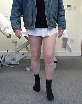
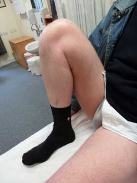
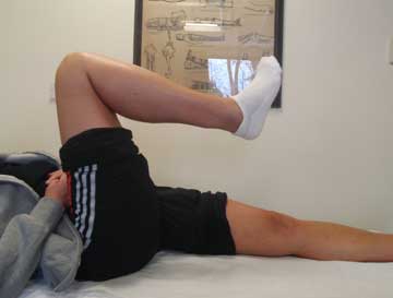
Like all operations prospective patients do not really know quite what to expect in the first months after osteotomy. Here are two examples of people who also had concerns about their osteotomy and the pictures give a good indication of the fact that you can walk well after the surgery and often quite quickly.
This photo is of a man who I reviewed recently in outpatients when he was 12 weeks after an osteotomy on his right side (left on the photo) and already extremely happy with his walking.
Previously he had ACL surgery on both sides, and he is a good example of someone who damaged his knee and had his ACL done but because of all the secondary damage his right knee was in valgus and he had to have an osteotomy on that side for the lateral compartment problem. So I went ahead with the osteotomy and put the right knee into varus.
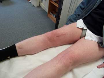
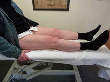
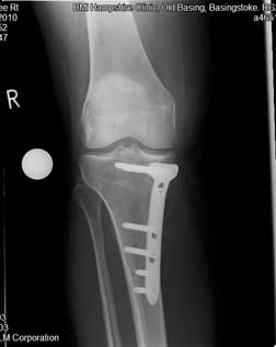
The second photo was taken twelve weeks after osteotomy
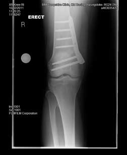
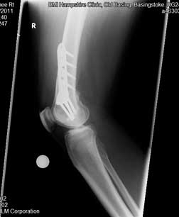
These are the two X-rays showing the femur with the plate in position from the front (left image) and the side (right image).
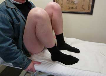
After the osteotomy, he was on crutches for six weeks protecting it because it was a femoral osteotomy (we protect femoral osteotomy with protected weight bearing on crutches for 6 weeks unlike tibial osteotomy where patients are allowed to fully weight bear without crutches from day 1 following surgery) . The ‘range-of-motion’ pictures were taken when he had been off his crutches for six weeks.
Finally, there is a video to show the patient walking.
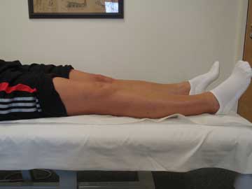
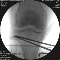
The next set of photos are of a young woman who has had a medial closing wedge osteotomy for lateral compartment disease only 3 weeks after the procedure was performed.
The image on the left is the fluoroscopic picture of the upper tibial region before the wedge was removed.
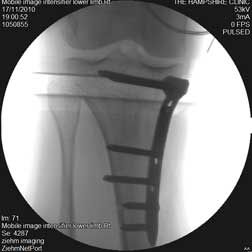
This last image shows the plate in place. Finally, there is a video to show the patient walking.
Full rehabilitation
It takes up to 12 months for the bones to fully heal. It is only after this time that we can contemplate removing the osteotomy plate.
Author
Professor Adrian Wilson
Knee Surgeon
Mr Adrian Wilson is a consultant knee surgeon based in London in the UK. His practice involves all aspects...read more
 First published 2010, and reviewed August 2023 by Dr Sheila Strover (Clinical Editor)