Knee anatomy includes all the various components of the knee, both inside and outside of the joint's waterproof capsule.
 Page updated March 2024 by Dr Sheila Strover (Clinical Editor)
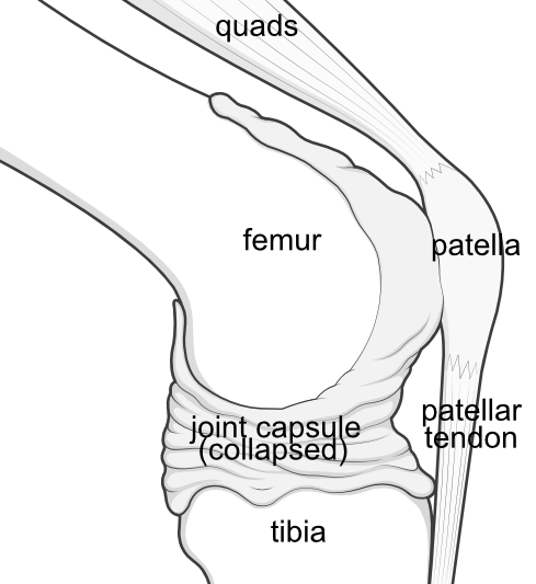
Side view - The knee is not a simple hinge but a fundamentally unstable moving joint of three bones - the femur (thighbone), tibia (shinbone) and patella (kneecap) enclosed on one end within a watertight capsule.
The rest of the knee's anatomical complexity relates to all the other structures that stabilise these bones while still allowing them to move.
Two other bones are involved in the knee, but are not part of this actual articulation - the fibula and the fabella.
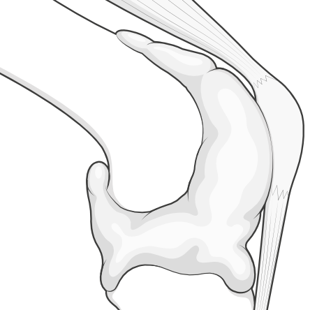
The capsule keeps the lubrication inside, where it is needed
This capsule is lined inside by a cell layer called the synovium which secretes the joint fluid that lubricates and nourishes the moving parts, and keeps the shiny, smooth articular cartilage joint surfaces healthy.
Damage to any structures inside the capsule may lead to an excess of this fluid, and possibly blood as well, causing the capsule to swell as the fluid cannot easily escape.
The stabilising cruciate ligaments are protected within a notch in the femur
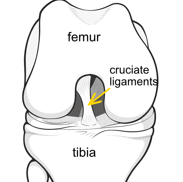
Front view of the knee (capsule removed for clarity).
The femur (thighbone) has two rounded knuckles at its lower end, and these are called 'condyles'. The rounded condyles make contact with the flattened top of the tibia and the undersurface of the patella.
Between the condyles is a notch, within which are contained the cruciate ligaments, important ligament stabilisers of the knee.
On either side of the joint, the femur and tibia bones are supported by the collateral ligaments (not illustrated).
Further stability and shock absorption is supplied by the meniscus
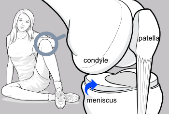 Oblique view - Sandwiched between the rounded condyles of the femur and the flattened top (plateau) of the tibia are the menisci (singular=meniscus) - shock absorbers that fill the space and further allow smooth movement of the joint. The menisci protect the delicate white articular cartilage at the ends of the bones from becoming damaged.
The one on the inner aspect of the knee is called the 'medial meniscus' and the one on the outer aspect of the knee is called the 'lateral meniscus'.
The patella is a fulcrum that facilitates extension
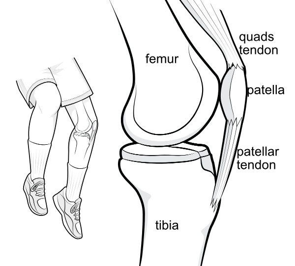
Outside of the capsule (not shown) the patella is tethered at its lower end to the tibia bone via the patellar tendon.
At its upper end the patella is attached to the quadriceps (quads) tendon, and thence to the quadriceps (quads) muscle. So contraction of the quads will force the leg straight (into extension).
It is kept in a central position by the walls of the trochlear groove in the femur.
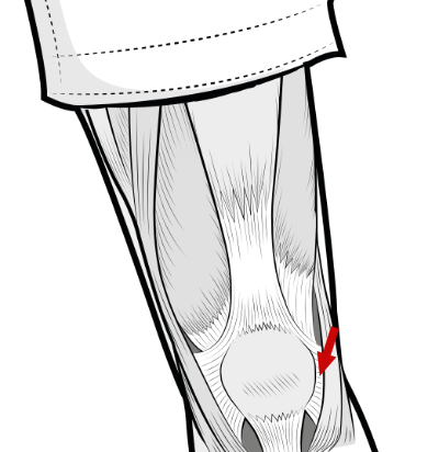
Besides being contained by the groove in the femur, the patella is further stabilised by a sheet of fibrous tissue on either side, including a strong band called the lateral retinaculum (arrow).
Patella, muscle and the fibrous tissue together are called the 'extensor mechanism' because they work together to support the knee when it is straightened (extended).
The muscles controlling flexion

At the back of the knee, contraction of the three hamstrings muscles of the thigh, together with gracilis, sartorius, plantaris, and popliteus, in concert with the calf muscle called gastrocnemius, cause the knee to bend.
When the thigh muscles contract the calf muscles relax, and vice versa.
Important nerves affecting the knee
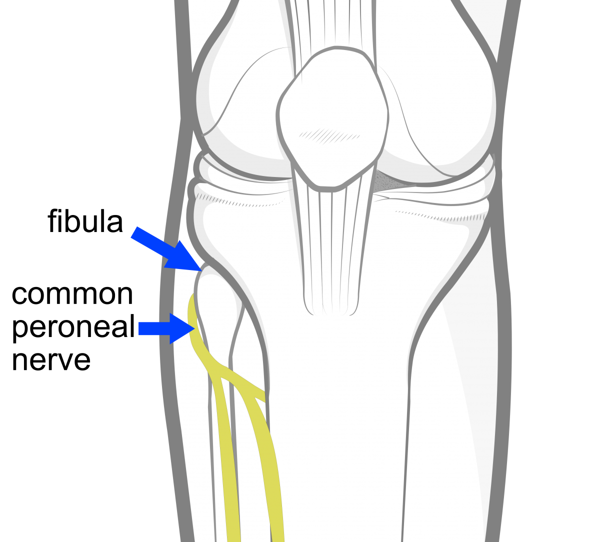
The sciatic nerve splits just above the knee into the tibial nerve and the common peroneal nerve. The other important nerve is the saphenous nerve.
Joint lining -
Bones -
Stabilisers -
- Meniscus
- Cruciate ligament
- Collateral ligament
- Anterolateral ligament
- Fat pad
- Lateral retinaculum
- Ligamentum mucosum
- Posteromedial corner
- Posterolateral corner
Muscles -
Nerves -
- Lateral femoral cutaneous nerve
- Saphenous nerve
- Superficial peroneal nerve
- Deep peroneal nerve
- Proximal tibial nerve
 2013 -