A suprapatellar plica is a fold of joint lining - possibly an embryonic remnant - that stretches across the joint cavity in the pouch above the kneecap.
 Page updated April 2024 by Dr Sheila Strover (Clinical Editor)
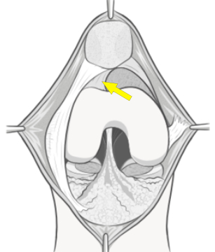
Here the patella (kneecap) has been cut from its tendon and lifted up to show the region at the top of the joint. You can see the curved suprapatellar plica above the patella.
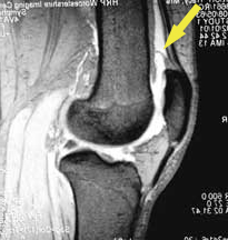
MRI image from the side showing the position (yellow arrow) of the suprapatellar plica. The plica may easily be missed on X-ray but may show up as a thin structure if there is fluid in the knee (which shows as white).
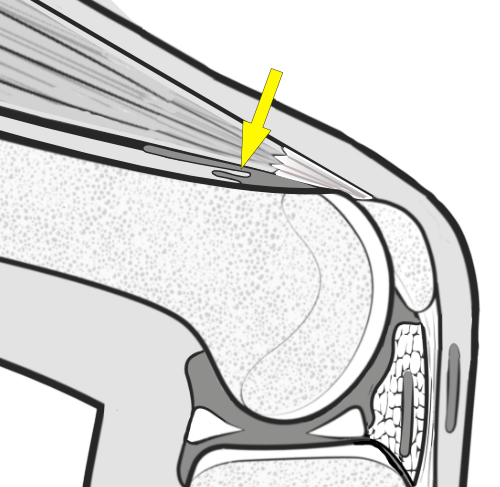
A traumatised and thickened suprapatellar plica can get trapped between the femur and the quadriceps tendon, where it can cause ongoing pain. Sometimes this is mistaken as quadriceps muscle or tendon injury.
Anatomy of the suprapatellar plica
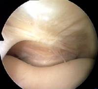
This arthroscopic photograph is looking down on the plica from above, and you can just see the top of the patella at the bottom of the image as it engages the groove of the femur.
Commonly a suprapatellar plica is a curved band along one wall of the capsule above the kneecap.
Rarely a suprapatellar plica may reach right across as a full membrane from one side of the supra patellar pouch to the other, dividing it in two, sometimes with a little window in the middle. Then it is called a suprapatellar septum. Although a septum can also be symptomatic, its real importance is that it often confuses the novice surgeon during arthroscopy.
-
Quote from peer-reviewed paper:
"....The knee joint is believed to have formed in the eighth week of fetal life with three compartments....Then, the synovial septa are partially resorbed over the next several weeks, and a single joint cavity is created...[and] remnants of the unabsorbed membrane are recognized as synovial plicae"
Citation: Akao M, Ikemoto T, Takata T, Kitamoto K, Deie M. Suprapatellar plica classification and suprapatellar plica syndrome. Asia Pac J Sports Med Arthrosc Rehabil Technol. 2019 Apr 22;17:10-15. doi: 10.1016/j.asmart.2019.03.001. PMID: 31044135; PMCID: PMC6477514.
-
Quote from peer-reviewed paper:
"Dull knee pain is a common symptom, and the pain is often aggravated during stair climbing...patients have tenderness over the upper edge of the patella. MRI examination is useful..."
Citation: Akao M, Ikemoto T, Takata T, Kitamoto K, Deie M. Suprapatellar plica classification and suprapatellar plica syndrome. Asia Pac J Sports Med Arthrosc Rehabil Technol. 2019 Apr 22;17:10-15. doi: 10.1016/j.asmart.2019.03.001. PMID: 31044135; PMCID: PMC6477514.
Management of a symptomatic suprapatellar plica
Often a suprapatellar plica becomes symptomatic only after a minor traumatic incident, because it is inflamed.
Conservative management, with rest and anti-inflammatory medication, may be enough to help it settle.
If symptoms become chronic, then the plica can be excised during arthroscopic surgery.
-
Quote from peer-reviewed paper
"In the presence of pathological plica associated with cartilage damage of the femoral condyle or patella at the time of diagnostic arthroscopy, plicae excision leads to favourable results in a high number of cases."
Citation: Zmerly H, Moscato M, Akkawi I. Management of suprapatellar synovial plica, a common cause of anterior knee pain: a clinical review. Acta Biomed. 2019 Dec 5;90(12-S):33-38. doi: 10.23750/abm.v90i11-S.8781. PMID: 31821281; PMCID: PMC7233704.
Relevant material -
eBook - An overview of plica problems in the knee
An expert look at the various plical folds of the knee and the problems that can be caused by a troublesome plica.
by Dr Sheila Strover (Clinical Editor)