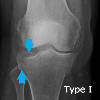
A tibial plateau fracture is a bone break through the flat top end of the shin bone (tibia).
 Page updated January 2024 by Dr Sheila Strover (Clinical Editor)
Why the tibial plateau is important
The tibial plateau is an important area because it is where the two menisci are seated and the two cruciate ligaments are tethered, both of which function as knee stabilisers.
Fractures or bone breaks of the tibial plateau not only have the potential to upset the delicate mechanics of the knee, but they are also frequently missed by the emergency team despite the patient complaining of severe pain in the knee following an injury.
One needs X-rays in two different planes to be able to see the often subtle outline of the fractured area. Also, the bone may not necessarily break off, but may be crushed and the fragment depressed, and it needs an experienced eye to appreciate this on X-ray.
What is the Schatzker classification?
Tibial plateau fractures are graded according to the Schatzker classification, so that anyone reviewing the patient's notes have an idea immediately of the severity of the problem.
Management of tibial plateau fractures
Tibial plateau fractures can be difficult to manage and cause problems with rehabilitation. Key issues include:
- assessing the degree of emergency
- restoring the anatomy
- avoiding residual stiffness from arthrofibrosis
- avoiding residual pain from arthritis
Assessing the degree of emergency - the injury may include damage to soft tissue structures as well as bone, including the menisci, nerves, blood vessels and ligaments. Some cases can be managed without surgery if the damage is not too severe. Very often, however, the initial management requires open surgery, and if there is vascular damage, including compartment syndrome, then immediate emergency surgery may be essential.
Restoring the anatomy - once attention has moved on from the immediate emergency, then the surgeon will attempt to evaluate the damage to the ligaments, especially the cruciate ligaments, and the degree to which any displacement of bone fragments is contributing to the functional damage. A CT scan may be called for so that the damage can be assessed in three dimensions, and the patient's bony injuries classified with the Schatzker classification for future reference. If there is no immediate emergency, blood in the joint (haemarthrosis) may be drained, and the limb put into a hinged brace until any surgery can be planned. Surgery might be undertaken in stages, trying to optimally align and fix in place the fractured pieces and restore ligament function.
Avoiding arthrofibrosis - in severe injuries like this, which may involve prologed periods of immobiisation and several surgeries, there is always a risk of residual stiffness from arthrofibrosis, and as much soft tissue mobility as is possible will become a focus as the bones unite.
Avoiding arthritis - long-term follow up will be necessary as there may be a strong possibility of future arthritis.
Quick links
Forum discussions
- brand new TPF
A physiotherapist ends up a patient after a nasty ski injury.
- TPF - 5 weeks out and backsliding - major ROM issues, help please
Patients discuss regaining range of motion after tibial plateau fracture.
- tibia plateau fracture, knee straightening
Patients talk about extension lag after tibial plateau fracture, and how to improve things with physiotherapy.
- Tibial plateau fracture
A patient's own story after fracturing the tibial plateau in a ski accident.
- manipulation
A discussion about managing stiffness after TPF.
Peer-reviewed papers
-
Quote:
"The crucial factor that influences long-term outcomes is the restoration of joint stability...The worst long-term outcomes were reported where there was associated ligamentous instability, meniscectomy, or alteration of the limb mechanical axis by more than 5 degrees."
Citation: Malik S, Herron T, Mabrouk A, et al. Tibial Plateau Fractures [Updated 2023 Apr 22]. In: StatPearls [Internet]. Treasure Island (FL): StatPearls Publishing; 2023 Jan-. Available from: https://www.ncbi.nlm.nih.gov/books/NBK470593/
-
Quote:
"The incidence rates of ligament injuries in tibial plateau fractures were as follows: Anterior Collateral Ligament (ACL) 26%, Posterior Collateral Ligament (PCL) 7%, MCL 24%, and LCL 14%. Medial collateral ligament injury was the most common in the Schatzker type 2 (50% of the injuries)."
Citation: Ebrahimzadeh MH, Birjandinejad A, Moradi A, Fathi Choghadeh M, Rezazadeh J, Omidi-Kashani F. Clinical instability of the knee and functional differences following tibial plateau fractures versus distal femoral fractures. Trauma Mon. 2015 Feb;20(1):e21635. doi: 10.5812/traumamon.21635. Epub 2015 Feb 2. PMID: 25825697; PMCID: PMC4362032.
Relevant material -
 2012 - Knee dislocation and tibial plateau fracture - a case presentation - by Professor Adrian Wilson (Knee Surgeon)
 2004 - Limb Realignment Procedure for Non-union of Tibial Plateau Fracture - by Dr (Mr) Mohi El Shazly (Knee Surgeon)
From the Journals -
- Journal interpretation - 2016 - Treatment strategy for tibial plateau fractures: an update - interpreted by Dr Sheila Strover (Clinical Editor)
