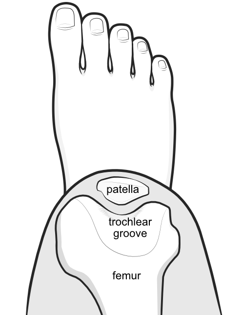
Patellar alignment is the position of the kneecap relative to the groove in the underlying femur bone into which it slides when the knee is bent and straightened.

The patella is normally V-shaped at the back and snugs into the V-shaped groove of the underlying femur.
What factors affect patellar alignment
Normally the articular cartilage at the back of the patella aligns perfectly with the articular cartilage in the underlying trochlea, which is a nice V-shape. The patella is normally held in this position by restraints from:
- the lateral side - lateral retinaculum
- the medial side - medial patellofemoral ligament (MPFL)
- below - the patellar tendon attached to the tibia bone via the tibial tubercle
- above - the quadriceps tendon of the quadriceps muscle which attaches at several places to the femur bone
- obliquely above - the vastus medialis obliquus part of the quadriceps muscle
- obliquely below - the medial and lateral retinacula
What factors cause patellar malalignment
Abnormal patellar alignment may affected by structural abnormalies patella or trochlea:
or by abnormal tension on the patella from other structures:
- Tight lateral retinaculum
- Tibial tubercle in poor alignment
- Torsion of femur or tibia
- Weak MPFL
- Weak quadriceps, especially VMO
Synonyms:
kneecap alignment
-