A 42-year-old female initially injured her knee skiing as a child at age 10.
She underwent surgical treatment of the knee with removal of a portion of the medial meniscus and was casted for a week at that time. Her treatment then left her with an injured knee that did not permit her to return to sports or normal activities of youth.
She recalled when we first met her that she had had pain for many years. She reinjured her knee at age 39 rollerblading. She underwent arthroscopy three months later for partial medial meniscectomy and removal of a loose osteochondral fragment. One week after surgery, she reinjured herself turning over in bed and withstood the pain for a year and a half before clinical evaluation.
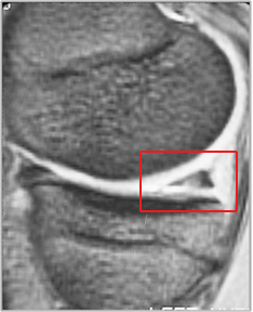
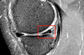
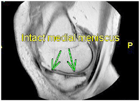
She presented to us with the MRI seen below. The red box on the MRI is over the medial meniscus cartilage. The MRI documents a chronic meniscus degenerative tear which had failed to respond to a partial meniscectomy. The options at this time when she presented to us were to repeat the meniscectomy. However, given the fact that it had already failed this did not seem to be a likely successful course of action. Therefore we planned to do a meniscus reconstruction where we would use a collagen scaffold as a regeneration template to regrow the missing meniscal tissue and therefore provide the shock absorber that her knee so clearly needed.
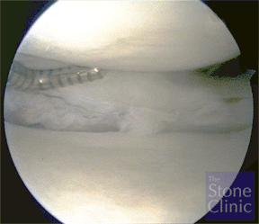
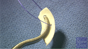
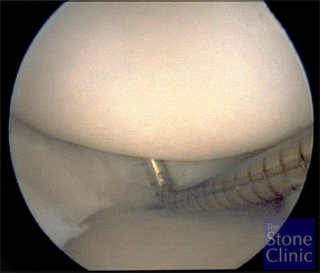
At the time of arthroscopy the meniscus was found to be chronically torn and degenerative as seen on the next picture. A meniscectomy if it had been performed at this time would leave her with essentially no meniscal rim and no protection for the joint – therefore a collagen meniscus implant was prepared as you can see in the photo on the right with a suture placed through the meniscus in order to help pull it into the knee after it was sized for the defect.