Then a beginner can dig deeper because of a clearer understanding of how the various components of the knee perform different roles.
The knee as a 'Hinge'
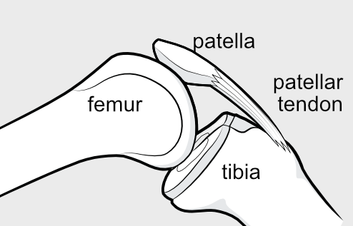 The two long bones of the knee are the femur (thighbone) and the tibia (shinbone). They are in contact with one another at the knee joint, allowing the knee to bend and straighten. Thus the knee is fundamentally a hinge, although some side-to-side and rotatory movement is possible. This hinge, however, is intrinsically unstable because the femur ends in two rounded surfaces, while the corresponding surfaces of the tibia are flat. This is why it is really an unstable hinge and other parts of the anatomy are needed in order to offer stability. A number of fibrous structures help to stabilise the joint, allowing the muscles around the knee to create the movement. The inside of the joint is lubricated with joint fluid and cushioning shock absorbers absorb any impact. All of these structures together allow fluid movement, but with stability and control, but damage to any of them might result in instability.
Ligaments improving Stability
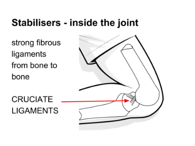
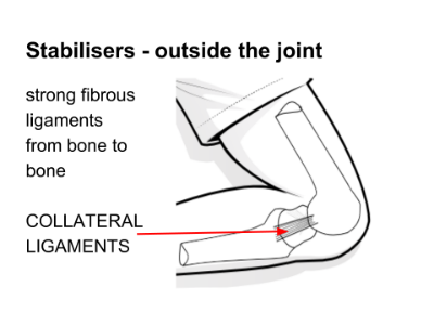
Key stabilisers are the cruciate ligaments and the collateral ligaments.
- Cruciate Ligaments - Right inside the joint cavity, in the middle of the knee, are two ligaments that cross over one another, preventing the two long bones from too much movement forwards or backwards in relation to one another. These are called the 'cruciate ligaments'.
Injury to these ligaments is dreaded by most competitive sportspeople, because they cannot heal themselves and surgery is technically challenging and rehabilitation is demanding. - Collateral Ligaments - Stabilising the joint in a side-to-side direction are another set of ligaments called the 'collateral ligaments'. The collateral ligaments are important, but healing occurs more readily and surgery is less often necessary.
Patella acting as a Fulcrum
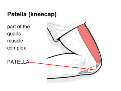
The patella (kneecap) is a bone actually embedded in the tendon of the quads muscle, and subject to the forces going through the muscle when it contracts. It serves as a kind of pivot for the hinge, and its lower end is fixed to the tibia bone via the bottom part of the tendon, which is known here as the patellar tendon or patellar ligament.
The patella and its relationship to the quadriceps (quads) muscle group is really important because it acts as a fulcrum, facilitating the quads movement and making the muscle contraction more efficient. As the knee bends and strightens, it is kept in alignment because it runs within a bony groove in the rounded end of the femur.
The menisci serving to absorb shock
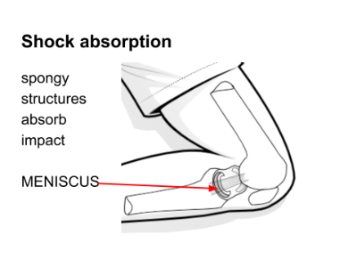
The menisci are cartilagenous structures tucked in between the femur and the tibia. Because the end of the femur is rounded and the end of the tibia is flat, a cushion-like structure called a meniscus fills the gap between the points of contact, to make the shapes more congruent and to absorb much of the impact.
A lot of the people who are researching online don’t know this term, because lay people tend to talk of the term ‘cartilage’ when they actually mean ‘meniscus’. The meniscus used to be called the ‘semi-lunar’ (or half-moon-shaped) cartilage, but this term has fallen into disuse in medicine, and it is now called the meniscus. What is called ‘cartilage’ is the white shiny gristle at the end of the long bones. Doctors talk down to patients sometimes and refer to the meniscus as the cartilage because they think that the patient understands that term better, but for the sake of researching online it is best to call the meniscus the meniscus, and to call the gristle at the end of the long bones the cartilage - and then there is no confusion.
Movement provided by muscles
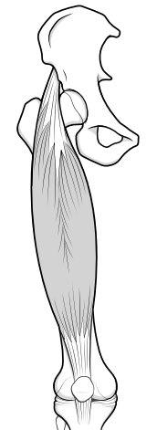
The muscles of the knee are several with the main muscle groups being the quads (or quadriceps) in the front of the knee and the hams (or hamstrings) at the back.
Because these muscles span the joint and are attached to the bone beyond it via their tendons, any contraction of the muscle creates movement at the hinge. Opposite muscles act as a team - with the one muscle group contracting, while the opposite muscle group plays out the tension and controls the overall movement.
All of these elements are important for normal knee movement, and injury to any one structure can disrupt stability and affect function. Because all the forces of the body are transmitted through the knee during normal sitting, walking and running even subtle changes in the anatomy can leave a person feeling pretty unhappy.
