Joint cartilage and meniscus damage may lead to alignment issues that can progress to arthritis.
 First published 2010, and reviewed August 2023 by Dr Sheila Strover (Clinical Editor)
Realignment osteotomy for knee pain - course
Part 1 - Introduction to the subject of knee osteotomy
- What is a knee osteotomy?
- An overview of the status quo in knee osteotomy
- Key concepts in knee osteotomy
- Knee osteotomy and painful osteoarthritis
- Opening wedge and closing wedge osteotomy
Part 2 - Osteotomy for varus and valgus deformity
- Indications for varus and valgus osteotomy
- Living with painful varus and valgus deformity
- High tibial osteotomy and distal femoral osteotomy
- Case study of high tibial osteotomy aiding ligament instability
- Benefits of varus/valgus osteotomy
- Potential problems with varus/valgus osteotomy
- Recovery post knee osteotomy
Part 3 - Osteotomy for patellar instability
When the layer of cartilage that lines the knee is damaged through wear and tear or trauma, osteoarthritis can develop.
This causes swelling, pain, stiffness and limitation of activity. If the damage to the cartilage is only slight– for instance a small loose flap – we as surgeons can perform a key-hole operation and tidy it up or carry out different types of cartilage surgery to treat the cartilage defect. If there is a significant problem with the joint surface cartilage, the key-hole options alone may be unable to improve matters. In cases like this case is the alignment of the limb needs to be optimal prior to carrying out major cartilage surgery.
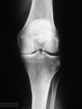
When you stand to have an X-ray taken of your knee, even though you are standing the X-ray will look as though there is thin air between the ends of the bones because the sandwich of the three non-bony structures - the joint surface cartilage on the end of the thigh, on the top of the shin and the meniscus inbetween - does not show up on the X-ray. If this sandwich is reduced by arthritis, the X-ray will reflect this as a loss of the "space" between the bones.
Osteoarthritis of the knee most commonly occurs in the inner (medial) compartment and frequently the rest of the knee remains relatively healthy. This X-ray shows this. To the left of the image you can see that the inner medial side is worn and has no space between the bones. The outer lateral side to the right of the image is normal with the gap taken up by with the 'invisible' joint surface cartilage and shock absorbing meniscal cartilage. (Remember that the lateral side is the side that has the skinny fibula bone next to the fatter tibia bone).
The most common scenario is that the patient presents to the surgeon with medial knee pain and are slightly knock-kneed (varus). When you do the long-leg x-rays to check the patient's alignment, the weight-bearing line is indeed going through the inner side of the knee. It is therefore possible to take the pressure off the inside of the knee, by re-aligning the leg and transferring those forces into the lateral side of the knee, which needs to be arthritis-free in order for the operation to work. This is a valg-ising osteotomy, as in the X-rays opposite.
The less common procedure is to do the reverse. This is done for patients with arthritis in their lateral compartment alone who are knock kneed/valgus. The surgeon can move the weight-bearing line over to the inner/medial side of the knee, which needs to be healthy for the operation to work.
This is a var-ising osteotomy.
Professor Adrian Wilson

Mr Adrian Wilson is a consultant knee surgeon based in London in the UK. His practice involves all aspects...read more