Only three bones make up the actual articulation of the knee - the femur, the tibia and the patella.

As the knee is bent, the two rounded ends (condyles) of the femur (thighbone) are exposed revealing the notch between them (intercondylar notch) as well as the cruciate ligaments which tether femur and tibia together. When the knee is straight, the condyles roll over and these structures are concealed. For the purposes of this illustration, the patella and its tendon have been omitted to allow you to see into the knee.
The knee surgeon performs many of the standard surgical procedures with the knee bent, creating the portals along the side of the patellar tendon for the instruments to enter this cavity.
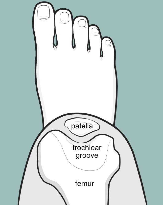
The patella and trochlear groove

The patella is a special bone because it is embedded in the tendon of the quadriceps muscle, and normally glides in the groove (trochlear groove) of the femur bone as the knee bends and straightens.
The lower part of the tendon is called the patellar tendon and is tethered below to the tibia bone. The upper part of the tendon is the quadriceps tendon and becomes the quadriceps muscle which is tethered to the femur.
Femur, tibia and intercondylar notch
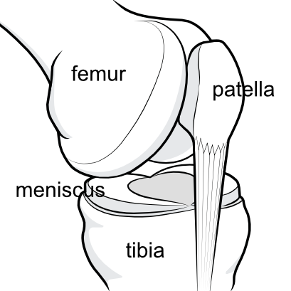
As one rounds the femur from top to bottom, the shallow trochlear groove becomes the deep intercondylar notch, where one can see the two cruciate ligaments tethering femur to tibia, and the two menisci sandwiched between the bones as a shock absorber.

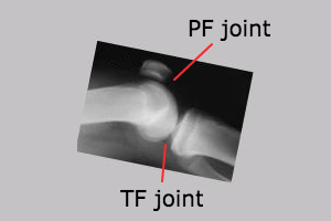
The knee is not a simple hinge - there are several bony points of contact. The contact surface between femur and tibia is called the tibio-femoral joint. Because the femur has two rounded ends, there is a medial and a lateral tibio-femoral contact area. The area of contact between the undersurface of the patella and the groove of the femur is called the patellofemoral joint.
Identifying the bones on X-ray
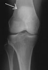
The white arrow is pointing to the outline of the patella, which is visible as a whiter circle 2-3 cm above the joint line.
The joint space between the bones appears as a black gap - actually it is not a gap but is filled with the menisci, which are not visible under x-ray. Compare the X-ray and the illustration to appreciate this. The joint space on X-ray should be about the same on both sides - if there is a difference (as in this film where one side is more closed than the other) then the assumption is made that there is some destruction of the meniscus on that side.
You can also see a thin bone on the outer (lateral) aspect of the tibia. This is the fibula. It is always useful to identify the fibula on an X-ray or drawing when you are trying to work out if you are looking at a right knee or a left one.
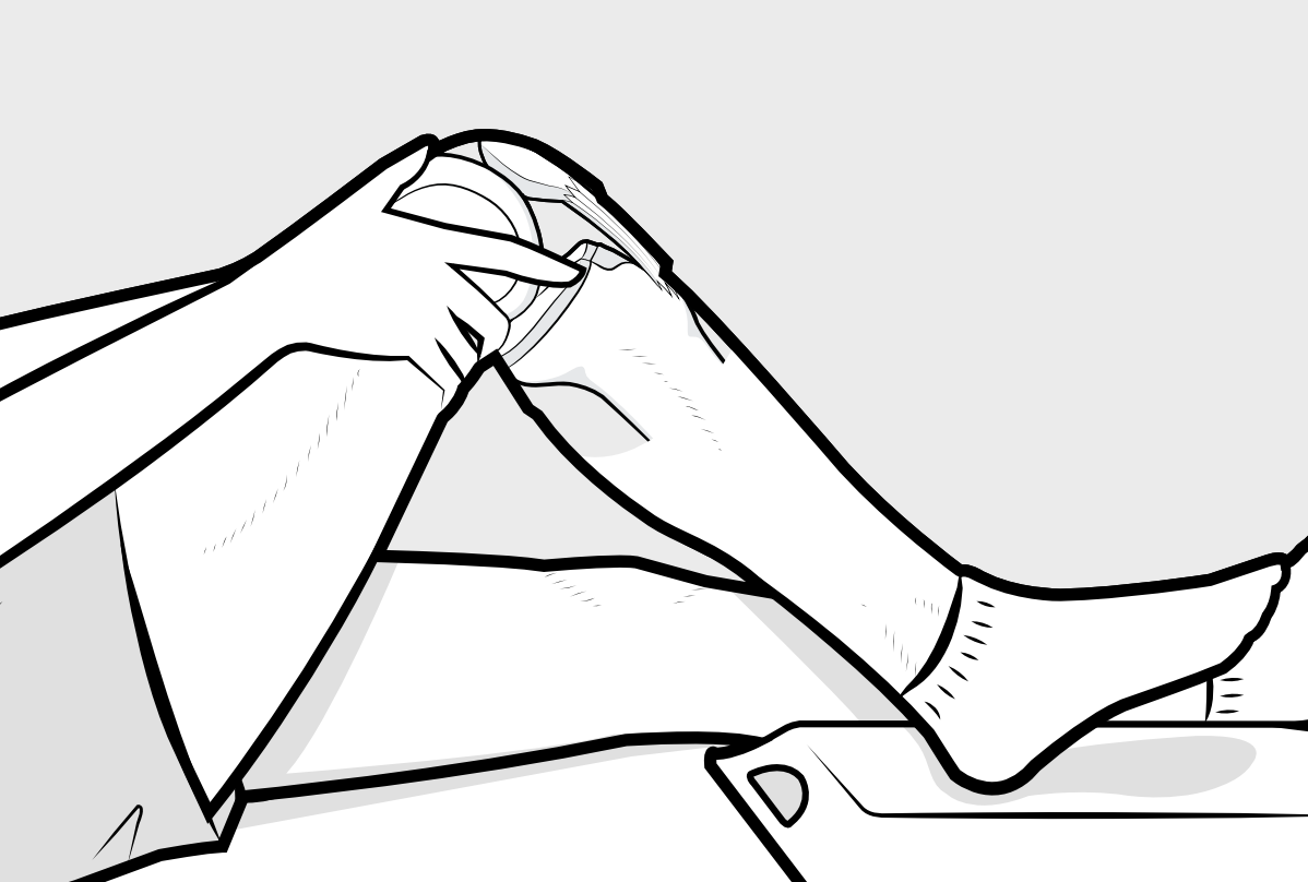

The position of the patella relative to the groove of the femur changes as the knee is bent. In the straight knee the patella lies above the groove and can be freely wiggled from side to side. As the knee is bent the patella engages with the groove of the femur and it should not be possible to wiggle it from side to side.
When you look at the X-ray again, you will see that the patella bone seems to be 'floating' - but it is just that the tendons above and below it do not show on X-ray. Neither do the two shock-absorber menisci, which appear just as a dark gap on X-ray between the two bigger bones.
Looking from above, when the knee is bent, the undersurface of the patella (kneecap) lies snugly like this in the trochlear groove. The patella should be central in the groove, and not favouring one or other side, and it should not be tilted. The sides of the patella and the walls of the groove should be almost parallel. The apparent gap that one sees on X-rays from this angle (Merchant views) is not really a gap but the space is filled with the white joint cartilage that covers both of the bony surfaces where they are in contact.

Note that you cannot see the rounded condyles of the femur in this view, nor can you see the tibia, as this angle looks down on the 'lap' and the condyles they are on the under-surface of the femur, while the tibia is at right angles to this view.
This view is also one that the surgeon can explore during arthroscopy, where he can get a superb opportunity to see how the patella glides in the trochlear groove. Many surgeons, unfortunately, miss this opportunity because they do not put the camera into this 'suprapatellar' region, and thus they fail to fully appreciate the anatomy here. This is also the best view to appreciate something called a 'plica', and problems with a plica may also be missed.
The tiny fabella
The fabella is a tiny bone which is not present in everyone, and mirrors the patella at the back of the knee.