Successful arthroscopic surgery dictates that the surgeon view the joint from more than one angle.
 First published by knee surgeon Angus Strover in 2008, and reviewed August 2023 by Dr Sheila Strover (Clinical Editor)
In this part of the course I am going to tell you why I have many happy patients who were unhappy with their previous arthroscopic surgery. And I am going to show you photos and videos.
I have published this to my colleagues in a leading surgical journals, but still today I have professional surgeon visitors who are amazed when I demonstrate this to them.
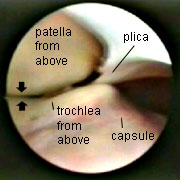
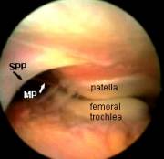
| Here is a drawing to demonstrate the view one obtains. Do you see how we are now able to clearly view the relationship of the undersurface of the patella to the underlying groove in the femur? From a suprapatellar portal one can best evaluate patellar tracking as the surgeon bends and straightens the knee. | 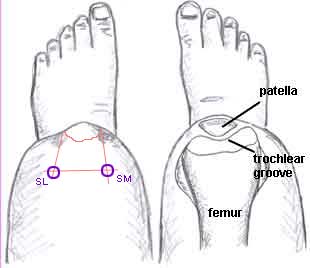 |
| As the knee is bent, the patella engages in the underlying trochlear groove of the femur. That thin fold of tissue, by the way, is a 'suprapatellar plica'. I will discuss this in a minute. These photos are of a right knee. |
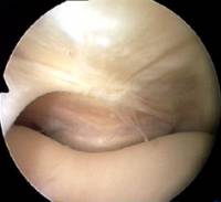 |
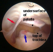
| As the knee is straightened, the patella lifts up out of the trochlear groove. |
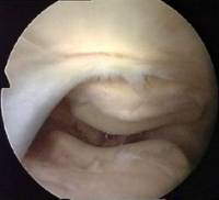 |
Medial Plicae
I've mentioned plicae already on this course. Although a plica is a normal structure, some people have them and some don't. They may differ in situation, size and thickness. If a medial plica is abnormal and thickened it can often be felt as a string-like object to the inner side (medial) of the patella. From the suprapatellar portal it is amazing how easy it is to see why medial plicae cause pain.
 |
 |
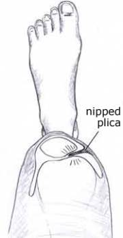
Look at this arthroscopic photo of a left knee. A plical fold is clearly seen being nipped between the patella and the femur. This is a medial plica, which is sweeping down out of view below the patella. The medial plica is found on the inner aspect of the knee. |
 |
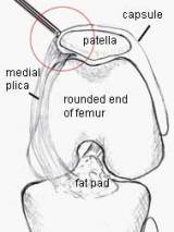 |
Look again at this region but this time from the front and side (all the left knee). |
Suprapatellar Plicae
The medial plica sweeps along the side of the patella, but the suprapatella plica lies horizontally above the patella,and may stretch right across the joint cavity (the 'suprapatellar pouch').
Here is an MRI scan, where the contrast has been reversed so that the bones are dark and the knee cavity light.
The yellow arrow is pointing to a suprapatellar plica, which you can see is stretched across the suprapatellar pouch above the patella.
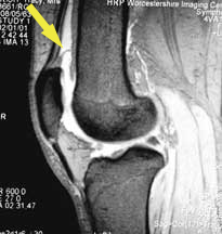
This is why one must measure two fingersbreadth above the patella before inserting the arthroscope, or the scope may be too low and this plica may be missed.
Here the arthroscope has been withdrawn above the level of the medial plica to reveal the suprapatellar plica (black arrow). You can see its position in relation to the medial plica (white arrow), which is just draping itself out of sight over the front of the femur. REMEMBER, WE ARE LOOKING FROM ABOVE, WITH THE SCOPE IS THE LARGE CAVITY ABOVE THE PATELLA. You can't see this structure from the ordinary portals!
Complete Septum and Fenestrated Plica
A suprapatellar plica may stretch right across the suprapatellar pouch, dividing it into two. This is called a 'complete septum', and its presence may confuse a novice arthroscopist. It is easier for you to understand a complete septum if you see an almost complete one - what we call a 'fenestrated' plica. That means 'a plica with a window'.
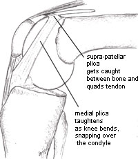
Bowstringing Plica
A plica may also be like a band of tissue. This image is of a suprapatellar plica viewed from the lateral suprapatellar portal, bowstringing and snapping across the supra patellar pouch over the medial femoral condyle. In this image, the red arrow is pointing to the plica. The little black arrows show the joint surface of the patella and femur.
The blue arrow shows the inflamed area of the femur where the plica has been snapping. This would be completely invisible from the lower portals.
Besides the advantages already mentioned, the supra-patellar portal allows -
- probing of the upper joint surfaces of the patella and the trochlea
- thorough examination of the lateral gutters
- examination of the fat pad
- examination of the anterior horns of the meniscus
I'm going to end this lesson at this point, but I just want to show you the video of patellar tracking. Click the arrow to start the video.
| Here are the stills for those of you who have trouble viewing the video. With the knee extended, the patella loses contact with the underlying trochlea, and is positioned above and slightly lateral to it. | |
| As flexion begins, the patella surface comes into contact with the trochlear surface. | |
| With further flexion, congruence increases. | |
| The patella is now in full contact with the trochlea. | |
| With the knee bent, the patella disappears from view over the trochlea. A suprapatellar plica can be seen in each photo. |